Referrers
Forgot your password
Complete the below form and one of our Medical Liaison team will be in contact to assist.
Referrer Support
At Wide Bay Nuclear Medicine, we are committed to supporting our referrers with accurate diagnostics, clear communication, and seamless collaboration. This page has been designed as a practical resource, providing information about our technology, referral processes, and how to connect with our Medical Liaison team. Our goal is to make referring simple, transparent, and supported at every step, so you can focus on delivering the best outcomes for your patients.
FAQs
What procedures are offered for Diagnostic Nuclear Medicine & Nuclear Medicine Therapy
Nuclear Medicine is the medical specialty that uses radiopharmaceuticals for diagnosis and therapy. The radiopharmaceutical is usually administered intravenously but, depending on the study, it may also be administered orally or by inhalation. The radiation dose from a diagnostic study is usually less than that of a CT scan.
Bone Scan
- Skeletal metastates
- Sports injuries (eg shin splints, stress fractures)
- Trauma (eg. scaphoid, sacral & femoral neck fractures not evident on X-Rays)
- Osteoporotic fractures
- Osteonecrosis (avascular necrosis)
- Osteomyelitis
- Painful prosthesis (loosening or infection)
- Back Pain – spondylolysis, pars interarticularis fracture
- Paget’s disease – presence & extent
- Enthesopathy, sacrolitis
- Reflex sympathetic dystrophy
Thyroid Study
- Evaluation of thyrotoxicosis
- Diagnosis of subacute thyroiditis
- Evaluation of functional status of thyroid nodules
Myocardial Perfusion: (“thallium”/”sestamibi”/”MPS”)
Assessment of myocardial ischaemia and viability. For patients unable to exercise dipyridamole or dobutamine (for asthmatics) are used. Rest and post stress images.
- Myocardial Perfusion Study (exercise / dipyridamole / dobutamine)
- Detection of presence, extent & severity of myocardial ischaemia
- Gated SPECT permits evaluation of LV wall motion & thickening & provides LVEF
Lung Scan
Ventilation and perfusion-pulmonary emboli. More sensitive and far less radiation than helical CTPA. Regional quantification. Right to left shunts.
Renal Scan (DTPA): (+/-captopril or frusemide)
Differential renal functions, renovascular hypertension, ureteric obstruction. MAG3 is substituted in children. Camera based GFR quantification.
Renal Study (MAG3 or DTPA) +/- Lasix
- Evaluation of renal perfusion and excretion
- Diagnosis of pelvi-ureteric junction obstruction & vesico-ureteric junction obstruction
- Follow-up evaluation after pyeloplasty
- Measurement of differential rela function
Renal Scan (DMSA)
Renal scarring and acute pyelonephritis (more sensitive than ultrasound). Differential function (particularly if one kidney is small or ectopic).
Renal Cortical Study (DMSA)
- ‘Gold standard’ test for detection of presence, location & extent of renal cortical scars
- Detection of acute pyelonephritis
- Measurement of differential renal function
Gallium Scan:
Detection of malignancy as a cause of PUO. Sarcoidosis. Chronic infections, eg osteomyelitis and infected joint prosthesis. Lymphoma staging and response to treatment.
Radioactive Iodine (1-131):
Treatment of hyperthyroidism.
White Cell Scan:
Localisation of acute or subacute infection. Assessment of activity of inflammatory bowel disease.
Colloid Liver Scan:
Diffuse or chronic liver disease. Assessment of liver nodules (FNH). Labelled Red Cell Scan Haemangioma of the liver. Gastrointestinal haemorrhage.
Biliary Study (HIDA):
Acute cholecystitis. Biliary obstruction or dysfunction. Response to a fatty meal or CCK used to assess chronic cholecystitis or sphincter of Oddi dysfunction.
- Chronic cholecystitis, gallbladder dyskinesia, cystic duct syndrome
- Acute cholecystits
- Post-operative biliary leak
Lymphoscintigraphy:
Diagnosis of lymphedema
Evaluation of regional lymphatic drainage prior to surgery (eg. for skin legions and breast carcinoma)
Sentinel node localisation in breast cancer and melanoma. Assessment of lymphoedema and lymphatic drainage.
Cerebral Perfusion Study(Ceretec):
- Dementias and cognitive impairment and cerebrovascular disease.
- Evaluation of dementia
- Localisation of focus of complex partial seizures
- Detection of presence and extent of cerebral infarction not evident on CT
Gastric Emptying:
Quantitative assessment of solid gastric emptying using a labelled egg sandwich. There are alternatives for patients allergic to eggs. Half clearance time and % retained at 4 hours are calculated. Liquid emptying can also be assessed (separate study).
Colonic Transit:
Small and large bowel clearance using sequential images over 5 days.
Lacrimal Scan:
Functional nasolacrimal duct obstruction.
Gated Heart Pool Study
- Evaluation of left & right ventricular size & systolic function, measurement of LVEF & diastolic filling parameters
Ventilation & Perfusion Lung Study
- Detection of pulmonary embolism
- Significantly less radiation than CTPA
- Preferred over CTPA for chronic pulmonary embolism
Parathyroid Study
- Localisation of parathyroid adenoma / hyperplasia in hyperparathroidism
Infection / Inflammation Study
- Both used to detect septic foci
Labelled White Cell
- Detection of active inflammatory bowel disease
GFR measurement (DTPA)
- GFR measurement
Captopril Renal Study
- Diagnosis of hemodynamically significant renal artery stenosis
Meckel’s Study
- Detection of Meckel’s diverticulum which contains ectopic gastric mucosa
Haemangioma Study
- Detection of cavernous haemangioma (eg. in liver)
Tumour Study
For tumour staging, evaluation of response to therapy, detection of residual or recurrent disease:
- Gallium – Lymphoma (HL and NHL), sarcoma
- MIBG (I-123 or I-131) – Phaeochromocytoma, neuroblastoma
- In-111 Pentetreotide – Carcinoid tumour, pancreatic islet cell tumours, medullary thyroid carcinoma, paraganglioma, glomus tumour
Gastrointestinal Transit Studies
- Oesophageal transit. Assessment of gastro-oesophageal reflux
- Gastric emptying. Utilising a labelled egg sandwich
- Colonic transit. Oral gallium-67 is monitored daily over 5 days
Therapy
- I-131 – Grave’s disease, toxic nodules, thyroid carcinoma
- Y-90 colloid – Radiation synovectomy (eg for RA)
- Samarium-153 EDTMP – Palliation of metastatic bone pain
- Strontium-89 – Palliation of metastatic bone pain
What equipment do you use at your Wide Bay Nuclear Medicine clinics?
Wide Bay Nuclear Medicine (WBNM) uses the latest technologies available to ensure patients receive the best possible care.
PET/CT camera
WBNM installed the first PET/CT camera in Hervey Bay in March 2020, the GE Discovery IQ.
SPECT/CT cameras
WBNM installed a GE Infinia™ Hawkeye® 4 (Hawkeye) in Bundaberg in 2006 and in Hervey Bay in 2007.
WBNM installed a GE NM 850 SPECT/CT camera in the new Bourbong Medical Centre in February 2020, and also in Hervey Bay in October 2021. Both cameras have now been upgraded to GE NM 860 SPECT/CT cameras, giving them the ability to perform diagnostic CT.
SPEC/CT provides WBNM with high-resolution scans for the most accurate diagnoses possible with a lower patient dose, because we understand that patient care cannot be compromised.
The following picture shows a series of images* taken by WBNM’s Hawkeye:
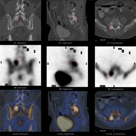
- The top row of images shows the low-dose CT scans in the coronal, sagittal and transverse planes.
- The middle row shows the corresponding nuclear SPECT images.
- The bottom row shows the fused SPECT/CT display.
The image series clearly demonstrates that the patient’s back pain was due to active facet joint arthritis, which WBNM’s Hawkeye accurately localised in the right side of the fifth lumbar vertebra. These precise images enabled WBNM to target specific treatment to the exact site.
* The image quality shown above has been reduced to allow the images to load quicker on the internet. The scans available to WBNM are of considerably higher resolution.
Dedicated Cardiac Gamma Camera
WBNM installed one of the first dedicated cardiac cameras in Australia in 2004, the IS2 Pulse™ – compact digital dual-head gamma camera. WBNM retired this camera in February 2020.
How we Support our valued Referrers
Our Medical Liaison Officers are here to assist with any concerns or requests, ensuring a seamless experience for referrers.
Our Team Can Help With:
- Ordering referral stationery and Medicare resources.
- Online image access.
- Connecting directly with a radiologist.
- Organising CPD events and referrer education sessions.
Contact Us:
Email: admin@widebaynm.com.au
Phone: (07) 4124 3580
Or complete the form below: